Cydar Maps
Automatic Volume Assessment
Revolutionizing Abdominal Aortic Aneurysm Monitoring

Find out more
Analyze
Automatic Volume Assessment
Cydar Maps Automatic Volume Assessment (AVA) is an essential component of our comprehensive solution, seamlessly integrating patient-specific features (e.g., anatomic vessel geometry) with clinical decisions (e.g., device selection) and patient outcomes (e.g., changes in aorta volume). This integration enhances clinical decision-making, providing a clearer understanding of patient progress and treatment efficacy.
Clinical Diagnostics
Post-EVAR Monitoring
Research & Development

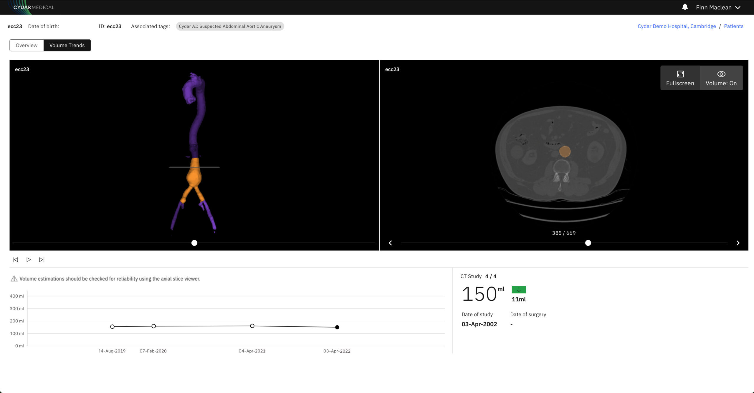
The Cydar Maps Automatic Volume Assessment (AVA) feature utilizes Artificial Intelligence (AI) to automatically estimate the volume of an Abdominal Aortic Aneurysm (AAA). Applied to any CT scan, AVA provides critical insights into potential changes in aneurysm volume over time, aiding in the assessment of the success of Endovascular Aneurysm Repair (EVAR) procedures. This innovative tool offers a streamlined approach to tracking disease progression.
Key features
Automatic Post-Op Assessment:
- Automatic assessment of post-op CT scans for AAA patients, including aortic aneurysm volume estimations.
- Volume calculation based on an assessment of the infra-renal aortic region.
Trend Visualization:
- Graphic display showing the trend in estimated volume calculations over time.
- Cydar Maps AVA tracks the progression of disease to inform clinical decision-making.
Objective Treatment Measures:
- AVA helps provide objective measures of EVAR treatment success, enhancing patient care (O’Donnell et al.).
Enhanced Communication:
- AVA can help share visualizations with patients and clinicians in a clear and understandable manner, fostering increased patient engagement.
Streamlined Monitoring:
- With AVA, surgical teams can streamline the follow-up monitoring process and reduce the time spent calculating aortic volume.
Benefits
Provides vital information to better inform clinical decisions regarding device selection and treatment strategies.
Enables timely interventions by detecting changes in aneurysm volume, potentially reducing the risk of complications.
Saves time and resources by automating volume calculations, allowing healthcare professionals to focus on patient care.

AI-driven assessments ensure consistent and reproducible results, reducing variability in manual measurements.
Secure, Compliant, Accessible

We are absolutely committed to the security of our platform and the protection of data.
Request a demo
A member of our team will be in contact with you to arrange an appointment. We look forward to meeting you.