Cydar Maps
Plan+ > Guide > Analyze
Surgical Planning
AI Enhanced Case Planning

Find out more
Plan+
Simplifying pre-op planning
This is aimed at providing precise, real-time navigation guidance, optimising patient outcomes and reducing surgical risks.

AI for vessel measurements

Guidewire and
Device placement

Highlight key points
of interest

Key features
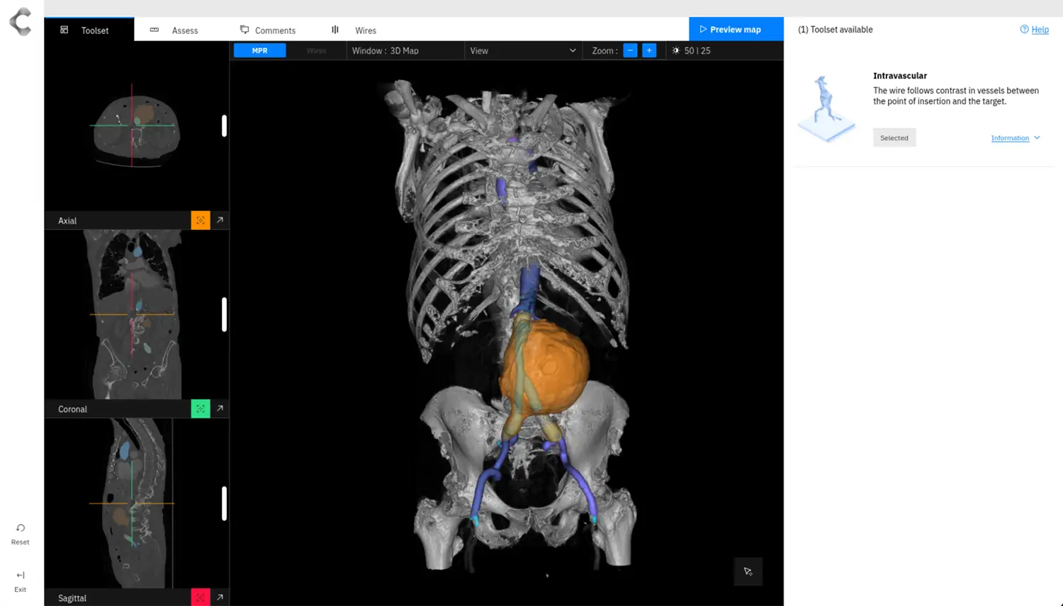
Multiple views:
- View AI segmentations of different vessel, skeletal and aneurysm structures.
- Toggle between Axial, Coronal, Sagittal and 3D map views.
- Ability to modify the AI segmentations further, utilising an extensive selection of manual tools, including vessel region growth and reduction.
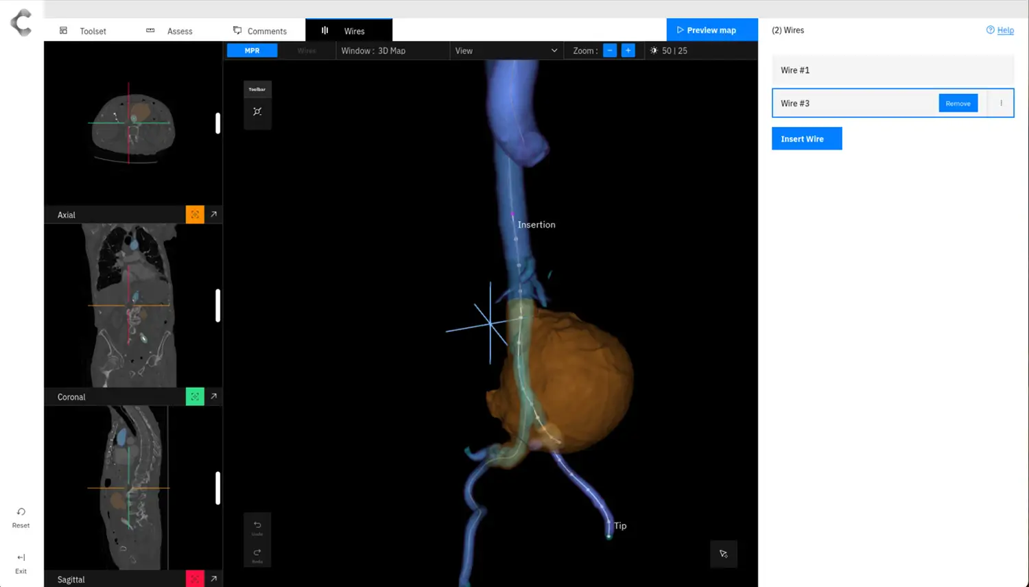
Wires:
- Semi-automatic insertion of multiple guide wires.
- Control and fine-tune positions.
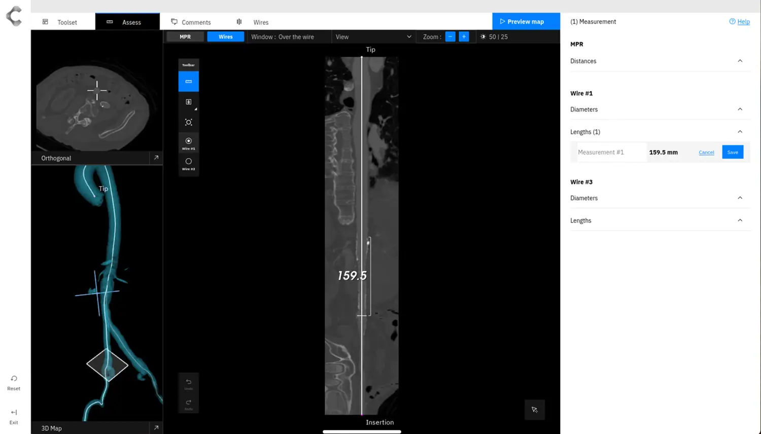
Measurements:
- Make dynamic measurements including Diameter, Length and Distance.
- Orthogonal and straightened workspace views provide additional geometric and spacial context.
- EVAR analysis through the Automated Aortic Volume Assessment tool.
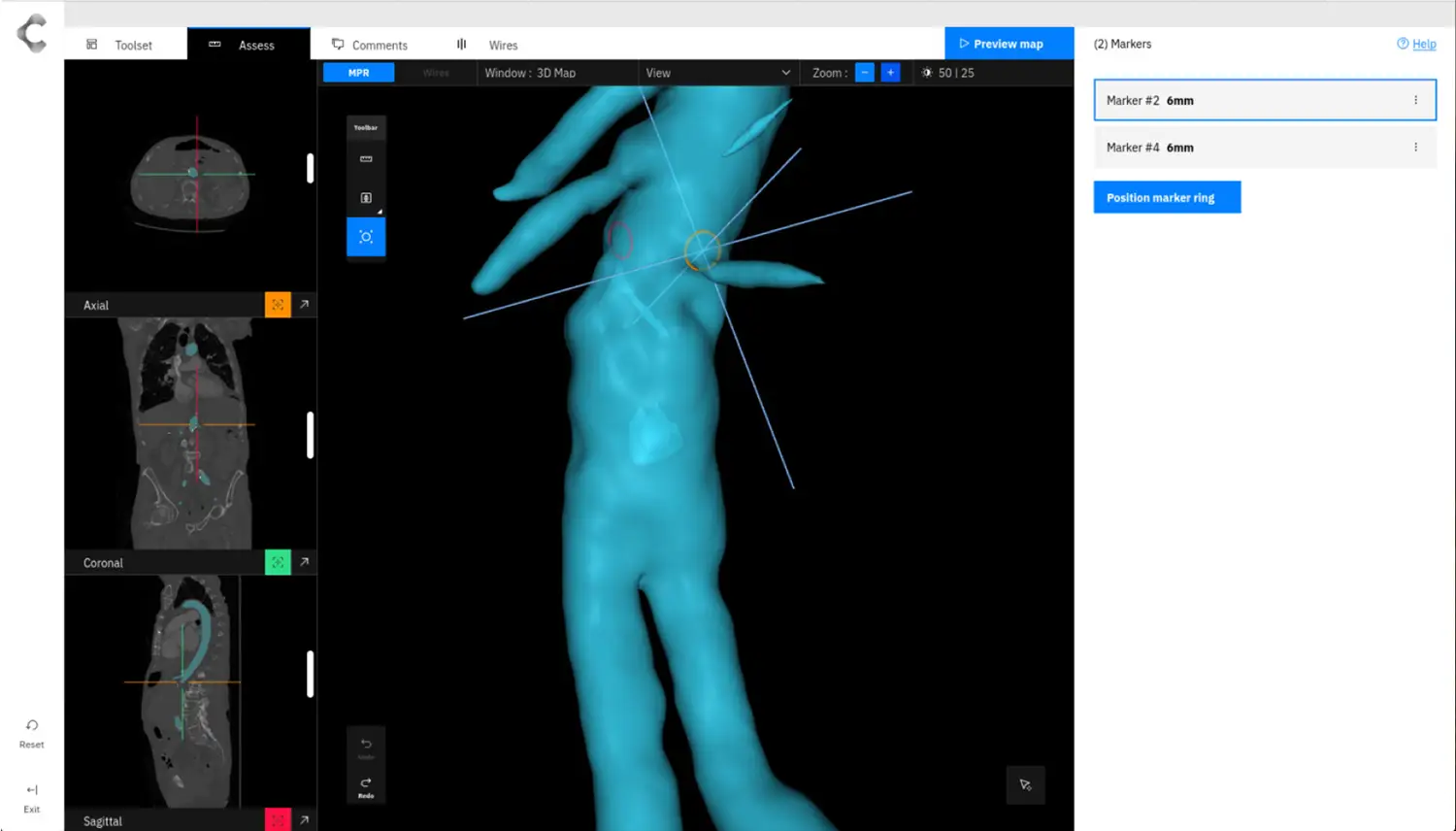
Marker rings:
- Assistance through automatic positioning of marker rings.
- Pin a marker ring, in one MPR view to lock it in place. Whilst manipulating the angle or position in another MPR view.
Commenting:
- Attach comments directly to measurements, marker rings and wires. Available to view at any point in time.
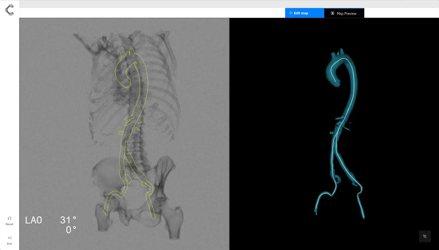
Preview Map:
- View your Map at point during the planning phase.
- The preview option provides a detailed understanding of the maps appearance in the OR.
Toolset:
- Additional functionality tailored to enhance specific surgical procedures, in the form of Toolsets.
- Navigate between different Toolsets to access these specific functionalities.
Benefits
A streamlined workflow that enables clinicians to plan a procedure with ease and efficiency. Generated Maps can be previewed within your Cydar eco-system.

Utilising Cydar’s cloud based technology empowers clinicians to create a map from any location from multiple device types.

Through Cydar’s ecosystem and collaborative features, maps can be shared for creation and consultation effortlessly.

Cydar’s AI segments the whole Aorta and Lumen. These can be viewed interchangeably to gain deeper context and facilitate better decision making.
Secure, Compliant, Accessible

We are absolutely committed to the security of our platform and the protection of data.
Request a demo
A member of our team will be in contact with you to arrange an appointment. We look forward to meeting you.